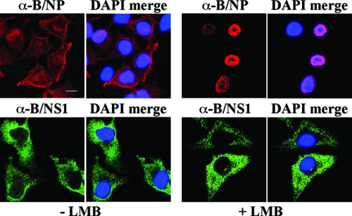
FIG. 9.
Cytoplasmic relocalization of the B/NS1 protein late in infection is not inhibited by LMB. MDCK cells were infected with influenza B WT virus. Starting at 3.5 h p.i., cells were left untreated or treated with LMB. The cells were fixed at 20 h p.i. and stained as indicated for the viral nucleoprotein (B/NP; top) or the B/NS1 protein (B/NS1; bottom) with primary monoclonal α-B/NP antibody or α-B/NS1 rabbit antiserum, together with secondary α-rabbit IgG-Alexa 488 or α-mouse IgG-Alexa 594 conjugate, respectively. DAPI was used to stain the nuclei. The cells were analyzed by CLSM, and the DAPI signals were analyzed by fluorescence microscopy. For each field, separate micrographs with the signal of the viral proteins alone (α-B/NP and α-B/NS1) and with merged DAPI signals (DAPI merge) are shown. Scale bar = 10 μm.

