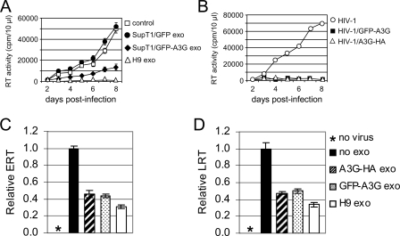
FIG. 11.
Exosomal A3G contributes to inhibition of HIV-1 replication. (A) Exosomes were isolated from culture medium of SupT1 cells transfected with GFP-A3G or GFP expression vectors. SupT1 cells were incubated without (control) or with indicated exosomes and then infected with vif-negative HIV-1. Additional doses of exosomes (10 μg/ml) were added 2, 3, and 5 days postinfection. Virus replication was monitored by RT activity released into the culture supernatants. (B) A3G tagged with HA or GFP retains full anti-HIV-1 activity when encapsidated into vif-negative virions. HIV-1 virions were produced by 293T cells transfected with vif-negative pNL4-3 plasmid DNA alone or in combination with expression vectors coding for GFP-A3G or A3G-HA. SupT1 cells were infected with equivalent amounts of viruses, and virus replication was monitored daily by RT activity released into the culture supernatants. (C and D) Exosomes secreted by 293T cells expressing A3G reduce accumulation of HIV-1 ERT (C) and LRT (D). SupT1 cells were preincubated for 16 h with 293T-derived exosomes or H9 exosomes or left untreated (no exo). Cells were infected for 2 h with wild-type NL4-3, washed, and cultured with an additional dose of exosomes for additional 6 h. Uninfected cells (no virus) served as a negative control. Relative levels of ERT and LRT were quantified by real-time PCR. Error bars, means ± SDs of triplicate samples.

