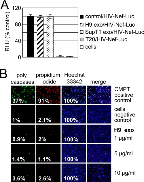
FIG. 8.
H9 exosomes do not inhibit HIV-1 entry or induce apoptosis. (A) Entry of HIV-1 into SupT1 cells was measured after 90 min of incubation of Nef-luciferase virus (HIV-Nef-Luc) with 1 × 10⁶ SupT1 cells preincubated for 16 h with exosomes or pretreated for 1 h with HIV-1 entry inhibitor T20 (1 μg/ml). Following incubation, cells were washed and incubated with cell-permeable luciferase substrate, and luciferase activity was measured in live cells. Luciferase activity in treated cells was normalized to luciferase activity of untreated cells (control) exposed to HIV-Nef-Luc (100%). RLU, relative light units; “cells” indicates untreated and uninfected cells. Error bars, mean values ± SDs of triplicate samples. (B) Activation of caspases and cell death in SupT1 cells exposed to H9 exosomes. Cells were left untreated (negative control) or were treated for 24 h with 10 μM camptothecin (CMPT) to induce apoptosis and cell death (positive control). Cells were treated with different doses of exosomes for 72 h followed by staining with FLICA (fluorescently labeled inhibitor of caspases) reagent (caspases) and subsequent staining with 1 μM Hoechst 33342 (nuclei) and 5 μM propidium iodide (dead cells). Fixed cells were observed under a fluorescence microscope using standard filter sets. Percentages of positively stained cells are shown.

