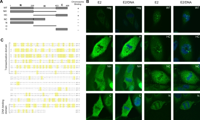
FIG. 2.
Both full-length and HC HPV-8 E2 proteins bind mitotic chromosomes. (A) Diagram showing the structure of wild-type (WT) HPV-8 E2 and the individual domains used for chromosome localization. N, H, and C represent the N-terminal, hinge, and C-terminal domains, respectively. The amino acid positions delineating these domains are shown. (B) Immunolocalization of HPV-8 E2 proteins and derived domains expressed in CV-1 cells. HPV-8 E2 proteins were detected with anti-FLAG antibody (green), and cellular DNA was counterstained with DAPI (blue). neg, negative. (C) Alignment of the amino acid sequences of the BPV-1 and HPV-8 E2 proteins.

