
FIG. 3.
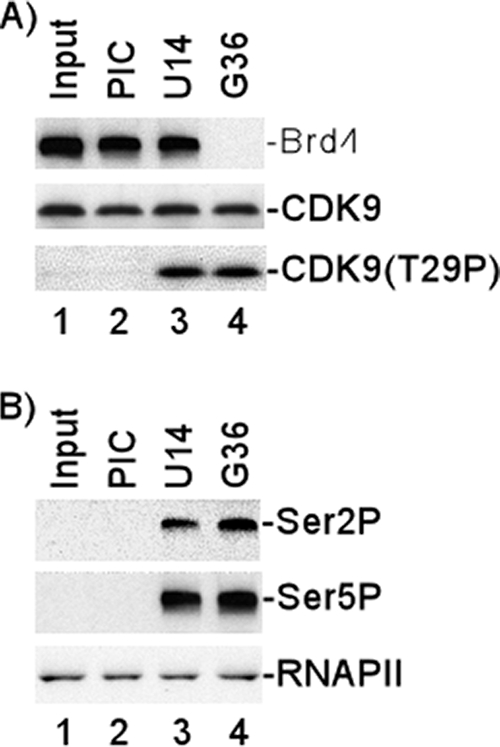
Association of Brd4 with HIV transcription complexes and CDK9 T29 phosphorylation during transcription. HIV-1 PICs were assembled by incubating biotinylated HIV-1 templates with HeLa nuclear extract and then purified with streptavidin-coated magnetic beads. The purified PICs were incubated with 50 μM ATP for 10 min and then washed extensively with 1× IVT buffer. The PICs were walked to position +U14 by incubation with 50 μM CTP, GTP, and UTP for 5 min at 30°C and then washed extensively with 1× IVT buffer. The TECs stalled at U14 were walked stepwise along the DNA by repeated incubation with different sets of three NTPs and then washed extensively with 1× IVT buffer to remove the unincorporated NTPs. (A) Protein compositions of PICs and TECs stalled at different stages were analyzed by fractionation by electrophoresis on 4-to-20% SDS-polyacrylamide gels, followed by Western blot analyses with antibody against Brd4, CDK9, or T29P CDK9. (B) Protein compositions of PICs and TECs stalled at different stages were analyzed by fractionation by electrophoresis on 4% SDS-polyacrylamide gels, followed by Western blot analyses with antibody against Ser 2P RNAP II CTD or Ser 5P RNAP II CTD. Western blot analysis with specific antibody (N-20) against the amino terminus of the largest subunit of RNAP II shown in bottom panel demonstrated equal amounts of RNAP II in the PICs and TECs.