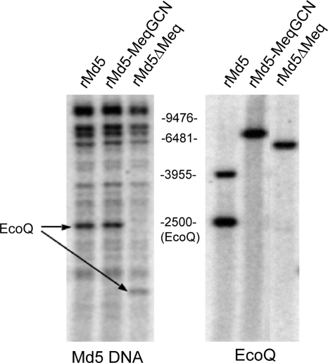
FIG. 7.
Southern blot analysis of rMd5, rMd5-MeqGCN, and rMd5ΔMeq. (A) DNA was digested with EcoRI and probed with total viral MDV DNA. The restriction profile of rMd5-MeqGCN is similar to rMd5, indicating that no gross genome rearrangements occurred. The arrow indicates the location of the EcoQ fragment. Due to the meq deletion in the EcoQ fragment of rMd5ΔMeq, this fragment migrates faster. (B) DNA was digested with PstI and probed with EcoQ fragment. The introduced LZ mutations in rMd5-MeqGCN resulted in the loss of a PstI site, and therefore a single band is observed, in contrast to the two bands for rMd5. Likewise, rMd5ΔMeq does not have a PstI site and, as a consequence of the meq deletion, results in a faster-migrating single band.

