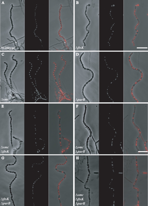
FIG. 2.
Confocal microscope images showing sporulation and DNA segregation phenotypes of the wild type and of single, double, and triple mutant strains. Four-day-old cultures grown on MS agar were sampled on coverslips, fixed, and stained with propidium iodide (red). In each panel, phenotypes of representative aerial hyphae are shown as phase-contrast and fluorescent image pairs, followed by a merged image. (A) M145 (wild type). (B) JM148 (ΔftsK). (C) HJ2 (Δsmc). (D) J2537 (ΔparB). (E) RMD3 (Δsmc ΔftsK). (F) RMD4 (Δsmc ΔparB). (G) RMD5 (ΔparB ΔftsK). (H) RMD6 (Δsmc ΔparB ΔftsK). Bar: 7 μm.

