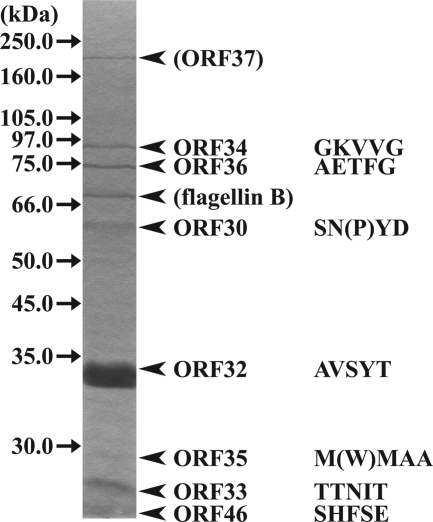
FIG. 2.
Identification of φRSB1 virion proteins. Proteins from purified φRSB1 particles were separated by sodium dodecyl sulfate-polyacrylamide gel electrophoresis (10% gel) and stained with Coomassie blue. The molecular size of each marker protein (Amersham full-range molecular weight markers and an LMW gel filtration calibration kit) is indicated on the left. The N-terminal amino acid sequence (five residues) determined for each φRSB1 protein band is shown on the right with its corresponding ORF. Amino acids in parentheses are obscure residues. Although the N-terminal sequence could not determined, the largest protein, approximately 175 to 180 kDa, is predicted to be ORF37, as there is no other candidate for this large size. A few small proteins were lost from the gel during electrophoresis.

