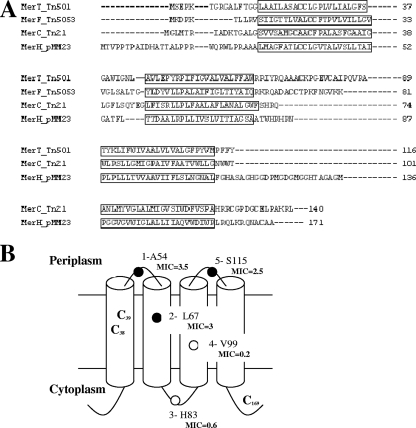
FIG. 2.
Sequence and topology of MerH. (A) Predicted protein sequence of MerH aligned with MerT, MerC, and MerF based on TM domain locations. Note that MerH has no significant similarity to the others at the primary sequence level. TM regions are shown in boxes and conserved cysteine residues in boldface type. (B) Topology of MerH determined by fusion to β-lactamase. Filled and open circles show fusions where the β-lactamase was found active and inactive, respectively. Carbenicillin MICs of strains expressing MerH-β-lactamase hybrid proteins are indicted in boldface type in mg ml−1.

