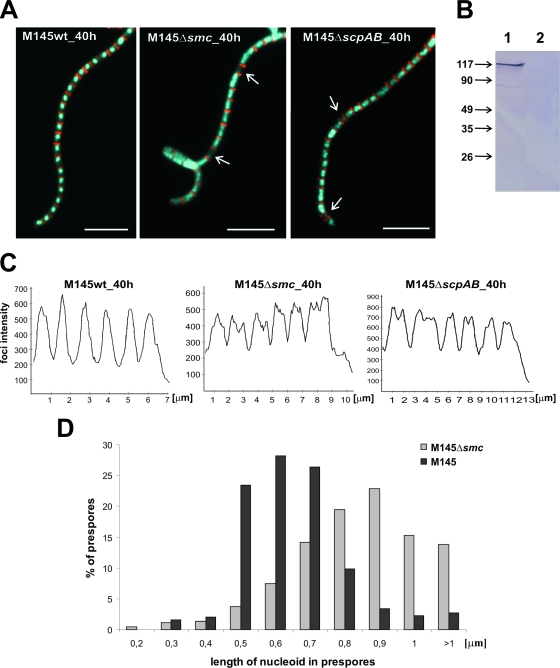
FIG. 1.
Influence of smc or scpAB deletion on chromosome condensation and segregation. (A) Examples of images showing hyphae with cell walls stained with WGA (red) and DNA stained with DAPI (blue). Anucleate compartments are indicated by white arrows. Scale bar, 5 μm. (B) Western blotting. Total proteins (20 μg) were separated by SDS-PAGE, and then SMC protein was identified by using antibodies raised against the recombinant SMC protein. Lanes: 1, wild type; 2, Δsmc mutant. Molecular sizes in kDa are shown on the left. (C) DNA signal intensity in spore compartments: fluorescent intensity of DNA stained with DAPI, presented in arbitrary units, was measured along a line drawn through the middle region of the spore compartments. (D) Percentage of prespores exhibiting different degrees of DNA compaction (length of nucleoid in prespores). wt, wild type; h, hour.

