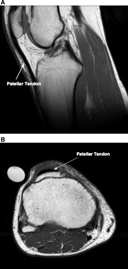
Fig. 1.
Representative sagittal [young man (23 yr old); A] and axial [older man (73 yr old); B] MRI images of the patellar tendon. The sagittal image is representative of a slice used to determined tendon length. The axial image is a representative slice from the distal portion of the patellar tendon.

