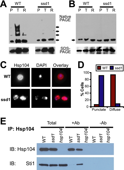
FIG. 7.
Ssd1 regulates Hsp104 function. (A and B) Native lysates prepared from wild-type (WT) or ssd1Δ cells after pretreatment (P), after treatment with the nonlethal heat shock (T), or after recovery for 1 h at 30°C (R) were analyzed by native PAGE (top) or by SDS-PAGE (bottom). The arrows in the top panel show the positions of Hsp104 migration on the native gels. Samples were prepared in the absence (A) or presence (B) of orthovanadate. (C) Differences between wild-type and ssd1Δ cells after nonlethal heat shock (37°C for 30 min and 46°C for 30 min) in Hsp104 localization were examined by immunofluorescence microscopy. (D) Cells in seven to eight independent microscopic fields (approximately 100 cells) were scored for the presence of a punctate or diffuse pattern of Hsp104. (E) Coimmunoprecipitation of Hsp104 with Sti1. Samples are total lysates (Total) and immunoprecipitation (IP) samples in the presence (Ab+) or absence (Ab−) of the Hsp104 monoclonal antibody (2B) from wild-type (WT), ssd1Δ, or hsp104Δ cells. Lysates were prepared from cells treated with nonlethal heat shock (37°C for 30 min and 46°C for 30 min). The samples were immunoblotted (IB) for Hsp104 or Sti1.

