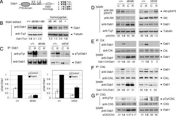
FIG. 1.
Effects of Dab1 phosphorylation site mutations on signal transduction. (A) Schematic of Dab1 protein structure showing the protein tyrosine-binding (PTB) domain and four tyrosine phosphorylation sites. (B) Expression level of Dab1 in embryonic brain. Protein extracts were prepared from E16.5 embryonic brains and analyzed directly by immunoblotting for Dab1 and tubulin. The samples on the left came from one litter, and those on the right were obtained from three different litters. The ratio of Dab1 to tubulin was calculated, with normalizing to +/+ brain extract. (C to G) Phosphorylation and binding events in Reelin-stimulated and control neurons. In each case, cortical neuron cultures were prepared from individual E16.5 embryos obtained from dab1ab/+ intercross or dab1cd/+ intercross timed matings. The embryos were then genotyped, and homozygous mutant and WT cultures were identified. Duplicate dishes were treated with control (C) or Reelin (R) for 15 min and then lysed. (C) Dab1 phosphorylation. Lysates were immunoprecipitated (IP) for Dab1 and immunoblotted for phosphotyrosine (4G10) and Dab1. Each graph shows means and ranges of the ratio of pDab1 to Dab1, normalized to the ratio in Reelin-stimulated dab1+/+ neurons, from two independent litters. (D) Akt phosphorylation. Lysates were analyzed using anti-Akt-pS473, anti-Akt, anti-Dab1 (B3), and Tuj1 anti-tubulin. The ratio of pS473Akt to Akt was calculated and normalized to the ratio in Reelin-stimulated dab1+/+ neurons. (E) Binding of CrkL to Dab1. CrkL immunoprecipitates were immunoblotted for Dab1 and CrkL. The lysates were also analyzed directly using Dab1 (B3). The ratio of Dab1 in the CrkL immunoprecipitate to total Dab1 was calculated. (F) Binding of Crk to Dab1 in neurons. The protocol is the same as that described above (E) except that Crk antibodies were used. (G) Phosphorylation of C3G in neurons. C3G immunoprecipitates were immunoblotted for anti-phosphotyrosine (4G10) and anti-C3G. The ratio of pC3G to C3G was calculated. Results in each panel are representative of two to three repeat experiments done using different litters of embryos.

