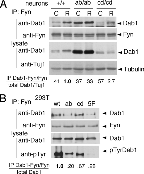
FIG. 2.
Dab1ab sites bind to the Fyn domain. (A) Neuron cultures prepared from WT, dab1ab/ab, and dab1cd/cd embryos at E16.5 were treated with control (C) or Reelin (R) for 15 min. Fyn was immunoprecipitated (IP) from lysates and immunoblotted for Dab1 and Fyn. The level of Dab1 in the Fyn immunoprecipitate was normalized for slight variations in Fyn immunoprecipitation. The level of Dab1 in the lysate was normalized for slight variations in loading using Tuj1 antibody as a control. The ratio of Dab1 in the Fyn immunoprecipitate to total Dab1 was then calculated and normalized to the ratio in Reelin-stimulated dab1+/+ neurons. (B) 293T cells were transfected with WT or mutant Dab1-green fluorescent protein (GFP) fusion proteins together with WT Fyn. Lysates were immunoprecipitated with anti-Fyn and immunoblotted with anti-Dab1 antibody. The level of Dab1 in the Fyn immunoprecipitate was normalized for slight variations in Fyn immunoprecipitation. The ratio of Dab1-GFP in the Fyn immunoprecipitate to total Dab1-GFP was then calculated and normalized to that of WT Dab1-GFP.

