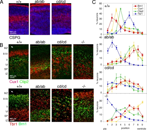
FIG. 4.
Abnormal preplate splitting and cortical lamination in dab1ab and dab1cd homozygous cortex. (A) E16.5 neocortex stained for CSPG (red) and DNA (blue). The subplate was intact in the WT but not in dab1ab or dab1cd mutants. (B) P1 cortex stained with Cux1 (red) and Ctip2 (green) or with Tbr1 (red) and Brn1 (green). The cortex was inverted in dab1ab/ab mice although not as severely as in dab1−/− mice. The dab1cd/cd phenotype was novel. Arrowheads point to the WT marginal zone, which is absent in the mutants. Scale bars in A and B are 400 μm. (C) Distributions of Tbr1+, Brn1+, Cux1+, and Ctip2+ cells in WT and mutant brains. Images were divided into nine equal-sized areas from the marginal zone (area 1) to the ventricular zone (area 9). The percentage of stained cells in each area was determined for each embryo. The graphs show means and standard errors of the results from three to four +/+, ab/ab, and cd/cd pups and two −/− pups.

