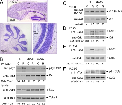
FIG. 5.
Intragenic complementation. (A) Normal development of cortex, hippocampus, and cerebellum of Dab1ab/cd mice. Brains (P20) were sectioned and stained with Nissl stain. Arrowheads indicate the normal migration of Purkinje cells. DG, dentate gyrus. (B to F) Phosphorylation and binding events in cultured neurons. dab1ab/+ and dab1cd/+ animals were crossed, and neuron cultures from individual E16.5 embryos were prepared. (B) Expression level and phosphorylation of Dab1 in neurons from dab1ab/cd and littermate dab1+/+ E16.5 embryos compared with dab1ab/ab embryos from another cross. Neuron cultures were treated with control (C) or Reelin (R) for 15 min. Cell lysates were immunoblotted with anti-Dab1 and anti-Tuj1 antibodies. The ratio of Dab1 to tubulin was calculated and normalized to the ratio in Reelin-stimulated dab1+/+ neurons. Lysates were also immunoprecipitated (IP) for Dab1 and immunoblotted with anti-phosphotyrosine (4G10) and Dab1 antibodies. The ratio of pDab1 to Dab1 was calculated and normalized to the ratio in Reelin-stimulated dab1+/+ neurons. (C) Activation of Akt by Reelin. Lysates were analyzed by using anti-Akt-pS473 and anti-Akt antibodies. The ratio of pAkt to Akt was calculated. (D) Binding of Crk to Dab1. Crk immunoprecipitates were immunoblotted for Dab1. The ratio of Dab1 to Crk in the immunoprecipitate was calculated. (E) Same as described above (D) but using CrkL antibodies. (F) Phosphorylation of C3G. C3G immunoprecipitates were immunoblotted with antibodies to phosphotyrosine (4G10) and C3G. The ratio of pC3G to C3G was calculated.

