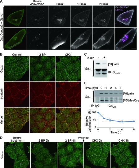
FIG. 1.
Dynamic palmitate turnover on Gαq/11. (A) Gαq shuttles between the PM and the Golgi apparatus. When Gαq-Dendra2, Gβ1, and Gγ2 were coexpressed in HeLa cells, Gαq-Dendra2 (green) was localized at the PM and the endomembrane. Gαq-Dendra2 in the endomembranes (upper) and a part of PM (lower) within the white regions was photoconverted by 405-nm laser. Converted Gαq-Dendra2 (gray scale) was monitored for 20 min. Cells were then immunostained with anti-GM130 (magenta) antibody (right). Scale bar, 10 μm. (B) Inhibition of palmitoylation causes detachment of Gαq/11 from the PM. HeLa cells were treated with 2-BP (100 μM) or CHX (20 μg/ml) for 4 h. The cells were then doubly stained with anti-Gαq/11 (green) and anti-β-catenin (red) antibodies. Scale bar, 20 μm. (C) 2-BP blocks palmitoylation of Gαq/11. HeLa cells were metabolically labeled with [3H]palmitate ([3H]palm) for 4 h either in the presence or absence of 2-BP. Gαq/11 was immunoisolated and subjected to fluorography (upper) or Western blotting (lower). IB, immunoblotting. (D) HeLa cells were treated with 2-BP for 4 h. Then, 2-BP was washed out, and protein synthesis was inhibited by 20 μg/ml CHX for 4 h. The cells were stained with anti-Gαq/11 antibody at the indicated times. The inhibition of palmitoylation for 4 h caused delocalization of Gαq/11 from the PM. The Gαq/11 dispersed by 2-BP came back to the PM again within 2 h after removal of 2-BP in the presence of CHX, indicating that the relocalization of Gαq/11 depends on palmitoylation and depalmitoylation. Scale bar, 20 μm. (E) Pulse-chase analysis of Gαq/11 palmitoylation. HeLa cells were labeled with [3H]palmitate or [35S]methionine-cysteine ([35S]Met/Cys) for 4 h. After incubation with chase medium for 0, 1, 2, 4, and 6 h, cells were lysed and subjected to IP with anti-Gαq/11 antibody. Immunoprecipitates were separated by SDS-PAGE, followed by fluorography. The ratio of [3H]palmitate to [35S]methionine-cysteine -labeled Gαq/11 was plotted in the graph. Error bars show ±SD (n = 3). IgG, control immunoglobulin G.

