FIG. 6.
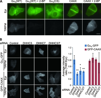
TIRFM imaging of membrane-bound palmitoylated Gαq. (A) HeLa cells were transfected with GFP-tagged Gαq (WT), palmitoylation-deficient Gαq (CS), or the C-terminal sequence of K-Ras including polybasic and prenylation sequences (GFP-CAAX). Cells were observed by epifluorescence microscopy (Epi) and TIRFM before and at 4 h after treatment with 2-BP. Gαq-GFP was clearly detected by TIRFM, and the intensity was reduced on 2-BP treatment. The intensity of Gαq (CS) was apparently weaker than that of Gαq (WT). Scale bar, 20 μm. (B) HeLa cells were transfected with control or DHHC3/DHHC7 siRNAs together with Gαq-GFP or GFP-CAAX. Scale bar, 20 μm. Relative fluorescence intensities of cell images from TIRFM compared to those of epifluorescence microscopy are indicated in the graph. Note that Gαq-GFP intensity visualized by TIRFM was reduced significantly in DHHC3 and DHHC7 knocked down cells. Error bars show ±SD (n = 5). **, P < 0.01; *, P < 0.05.

