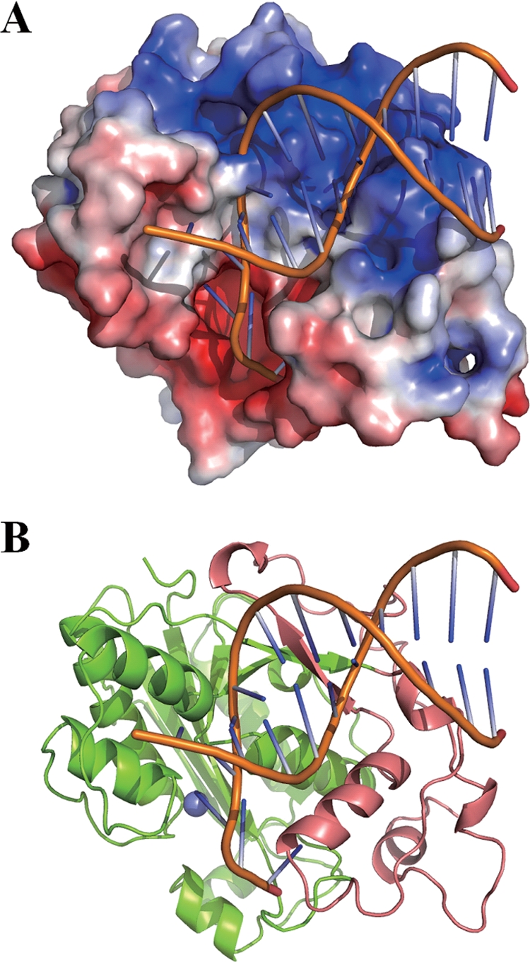
FIG. 7.

Structural model of CRN-4 bound with a DNA. (A) A model of a 12-mer dsDNA (PDB entry 1KLN) bound at the basic Zn-domain of CRN-4 was constructed by using Zdock (http://zdock.bu.edu/). The two phosphate backbones of DNA are bound snugly in the two cleft regions of the Zn-domain. For clarity, only one subunit of CRN-4 in the dimer is displayed. (B) The ribbon model of CRN-4 demonstrates that DNA is bound at the Zn-domain (in salmon) and further extended to the DEDDh domain (in green) for cleavage. The Mn2+ bound at the active site is displayed as a blue ball.
