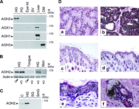
FIG. 2.
Tissue- and cell-specific expression of the AOH2 protein and mRNA. (A, B, and C) Tissue and organ distribution of the AOH2 protein. Extracts (100 μg) of the indicated tissues and organs were loaded on an 8% polyacrylamide gel run in denaturing and reducing conditions, transferred to nitrocellulose membranes, and subjected to Western blot analysis with anti-AOH2, anti-AOX1, anti-AOH1, anti-AOH3, and anti-β-actin antibodies. Abbreviations: PG, preputial gland; SV, seminal vesicles; OM, olfactory mucosa; SG, salivary gland; SmG, submaxillary gland; C = extracts of HEK293 cells transfected with the full-length AOH2 cDNA used as a positive control for the experiment. (D) Tissue sections derived from HGs (a and b) and skin (c to f) were hybridized to a 35S-radiolabeled AOH2 sense (a and c) or antisense (b, d, e, and f) riboprobes. Magnifications, ×200 (a and b), ×100 (c and d), and ×400 (e and f). Abbreviations: sbl, suprabasal layer; bl, basal layer; sg, sebaceous glands; M, molecular mass.

