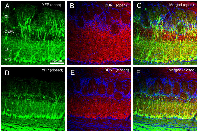
Figure 3.
Representative double-color BDNF immunocytochemistry of the olfactory bulb of thy1 YFP transgenic mice (YFP) following 30 days of sensory deprivation as imaged on a confocal system. (A) Contralateral control OB (YFP (open)). (B) Same, but demonstrating BDNF-ir (BDNF (open)), and (C) merged image. (D–F) Same as (A–C) but for the OB ipsilateral to the naris occlusion. EPL, external plexiform layer; GL, glomerular layer; MCL, mitral cell layer; Scale bar, 100 μm.

