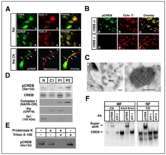
FIGURE 1. Identification of mitochondria as a site of CREB localization in neurons.
A, both CREB (panels a– c) and pCREB (Ser-133) (panels d–i) colocalize with mitochondria in the cerebral cortex of rat adult brain in situ (panels a–f) and primary cultured cortical neurons (panels g–i). White arrows indicate colocalization with punctate staining of pCREB and mitochondria in a single cortical neuron. B, pCREB immunoreactivity (panels a and d) with cytochrome c (Cyto. c) (panels b and e) is impaired in dorsal root ganglia in CREB null (−/−) mice (panels a– c) but not in CREB+/− (panels d–f). C, immunogold electron microscopy shows pCREB in the mitochondrial matrix of cortical neurons. Mito, mitochondria; Nu, nucleus. Scale bar: 200 nm. D, CREB is found in the mitochondrial subcellular fraction. pCREB was detected as a 43-kDa band. The same blot was then stripped and reblotted with anti-complex I (mitochondrial NADH oxidoreductase (OR), 37-kDa subuint) (mitochondrial marker), Bip (GRP78) (endoplasmic reticulum marker), and Sp1 (nucleus marker), each of which were also detected. N, nuclear fraction; C1, cytoplasmic fraction 1; P1, pellet 1 mitochondria-rich fraction; P2, pellet 2 mitochondria fraction. E, proteinase K assay using the mitochondrial fraction shows that pCREB is localized to the mitochondrial matrix. F, mitochondrial fraction (MF) of cortical neurons (CN) and adult rat brain show specific CREB DNA binding activity by EMSA. EMSA was performed with mitochondrial and nuclear extracts using 32P-labeled CRE oligonucleotides. Supershift analysis for identification of CREB complex used antibodies (Ab) for pCREB (Ser-133), ATF-1, and ATF-2. NF, nuclear fraction.

