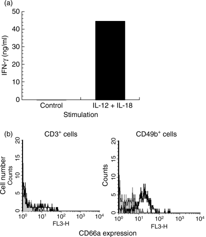
Figure 4.
CD66a expression by in vitro activated natural killer (NK) cells. (a) Interferon-γ (IFN-γ) was measured by enzyme-linked immunosorbent assay in the supernatant of 3 × 10⁶ BALB/c spleen cells incubated in the absence or in the presence of interleukin-12 (IL-12) and IL-18. (b) CD66a expression, analysed by flow cytometry, by CD3+ and CD49b+ BALB/c spleen cells cultured for 3 days in the presence of IL-12 and IL-18. For each cell population identified with specific marker, the grey zone represents background fluorescence, the bold line indicates labelling with CC1 anti-CD66a antibody.

