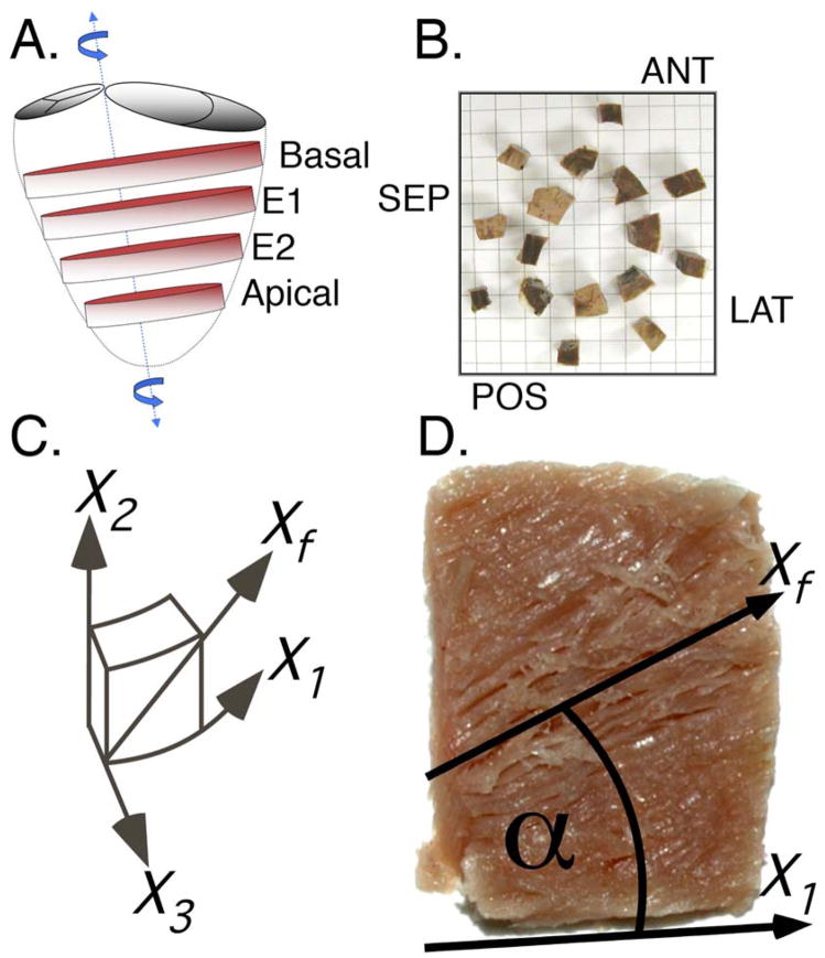
Figure 1.
Each heart was systematically sectioned for quantitative histologic analysis of the myofiber helix angle. (A) Basal, mid-equatorial (E1 and E2), and apical slices were sliced out of each heart. (B) Each slice was then sectioned into sixteen tissue blocks and eight anatomical sectors (numbered) were used for histologic analysis. Eight chunks of remaining tissue (not numbered) are shown in approximate anatomical location, viewed from base to apex. (C) A “cardiac” coordinate system was defined for each block to enable calculation of the myofiber angle (X1–circumferential, X2–longitudinal, X3–radial). (D) The myofiber angle for each transmural section was measured using computer software and was referenced to the local X1 axis.

