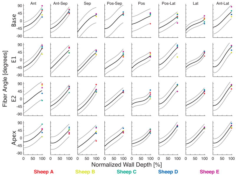
Figure 2.
Transmural myofiber angles in each of the 32 anatomical sectors, color-coded for each heart. The cubic fit is shown in each sector as a black line, with 95% confidence intervals in gray. The anterior papillary muscle was located between the anterior and anterior-lateral sectors and the posterior papillary muscle was located between the posterior and posterior-lateral sectors.

