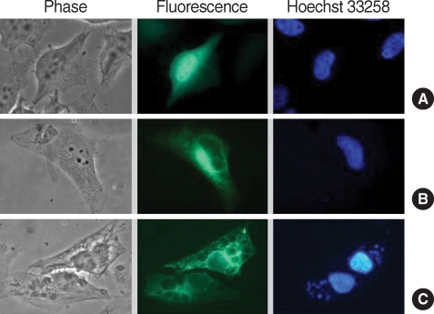
Fig. 3.
Localization of GFP-CAMLG fusion protein expressed in HeLa cells infected with T. gondii. (A) control of pEGFP-N1 plasmid only; (B) expression of pEGFP-CAMLG plasmid near nucleus; and (C) expression of pEGFP-CAMLG plasmid near nucleus spreads to the PVM after T. gondii infection. In each set green color indicated the fluorescence of GFP chimera and blue color the fluorescence of Hoechst 33258 from nucleus of the cell or T. gondii.

