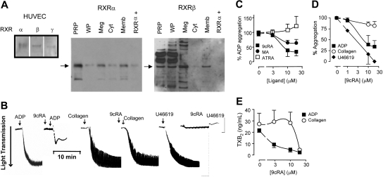
Figure 1.
Human platelets express functional retinoid X receptors. (A) Western blot analysis showing positive controls for RXR isoforms (α, β, and γ) in HUVECs, and the expression of RXRα and RXRβ in human platelet-rich plasma (PRP), washed platelets (WPs), megakaryoblast cell line Meg-01 (Meg), and the cytosol (Cyt) and membrane (Memb) fractions of PRP. In addition, a positive control peptide for RXRα (RXRα+) was also included. (B) Typical aggregometer traces showing the changes in light transmittance seen as platelets aggregate (in sequence from left to right) to ADP (4 μM; left panel), 9cRA (10μM) given 3 minutes prior to ADP; to collagen (1 μM), 9cRA (10 μM) given 3 minutes prior to collagen; and to the TXA2 mimetic U46619 (1 μM) and 9cRA (10μM) given 3 minutes prior to U46619. (C) RXR ligands 9cRA (■), and methoprene acid (MA; ●), but not the retinoic acid receptor ligand all-trans retinoic acid (ATRA; □) inhibit ADP-induced platelet aggregation. Figure represents the mean ± SEM changes in percent of ADP aggregation. (D) 9cRA (1-20 μM; 3-minute pretreatment) inhibits ADP-induced (2 or 4 μM; ■) and U46619-induced (1 μM; ♦), but not collagen-induced (1 μM; ○) platelet aggregation in a concentration-dependent manner. Figure represents the mean ± SEM changes in agonist-induced aggregation (titrated to approximately 85% of maximum). (E) 9cRA (1-20 μM; 3-minute pretreatment) more potently inhibits ADP-induced (2 or 4 μM; ■) and collagen-induced (1 μM; ○) platelet TXA2 secretion. Released TXA2 was measured in PRP at the end of aggregation assays (15 minutes) by using an assay for its stable metabolite, TXB2. Figure represents the mean ± SEM TXA2 release. Data represents result from at least 4 separate donors.

