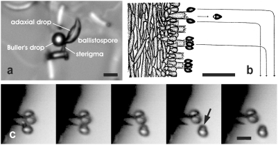
Figure 1. The process of ballistospore discharge.
a, Ballistospore of Tilletia caries a few seconds before discharge. b, Predicted trajectories of spores discharged from a mushroom gill illustrated by A. H. R. Buller [1]. c, Successive images of ballistospore discharge in Armillaria tabescens from video recording obtained at 50,000 fps. Buller's drop (arrowed) carried with discharged spore. Scale bars, a, c, 10 µm b, 50 µm.

