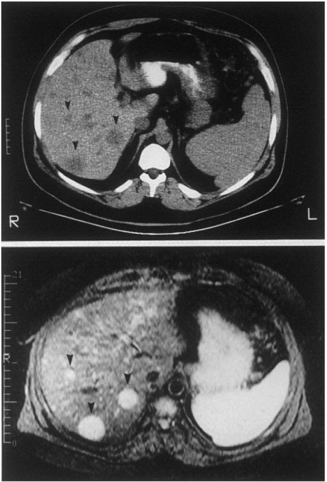
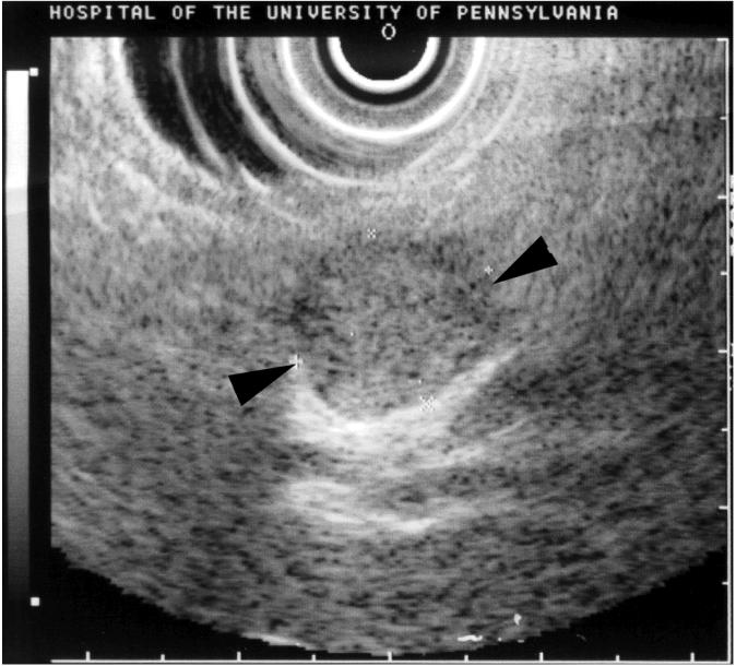
Figure 2.
CT, MRI, EUS in patients with PETs. Panel A illustrates CT (top) and MRI(bottom) images of the abdomen in a patient with a metastatic gastrinoma. Liver metastases are indicated by arrowheads. Panel B illustrates an endoscopic ultrasound image of a pancreatic body insulinoma confirmed at subsequent surgery. The tumor is indicated by the black arrowheads.


