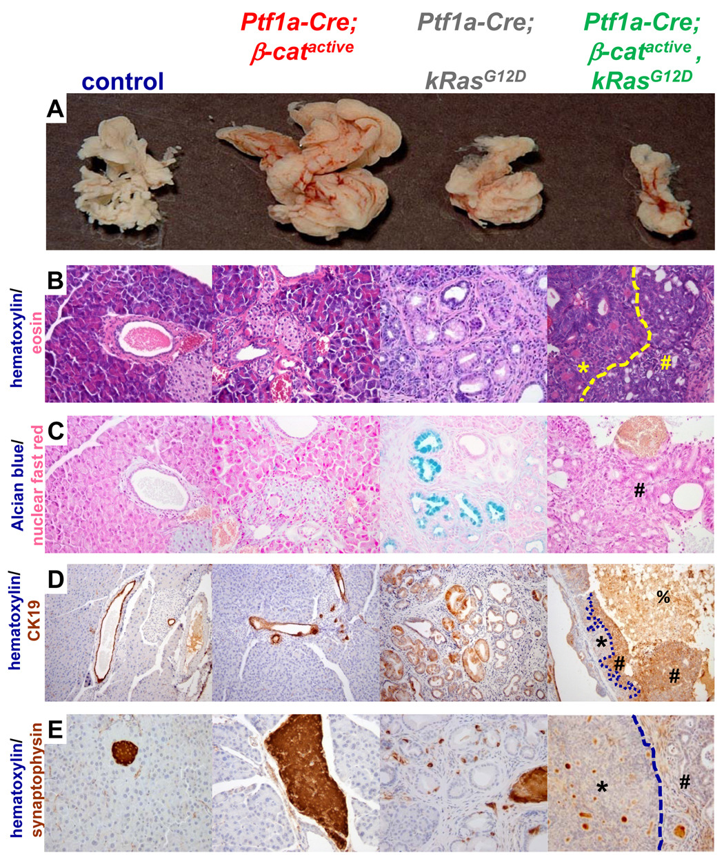
Figure 6. PanIN lesions do not form in Ptf1a-Cre; β-catactive, KrasG12D mice.
(A–E) 3 month old pancreata. (A) Pancreas size is reduced and morphology is condensed in Ptf1a-Cre; β-catactive, KrasG12D mice when compared to control or Ptf1a-Cre; KrasG12D organs. Ptf1a-Cre; β-catactive mice exhibit pancreatic hyperplasia in this mouse background that is equivalent to what was previously described. (B) Hematoxylin (blue)/eosin (pink) pancreas sections, 400x magnification. Two distinct tumor types that frequently form within Ptf1a-Cre; β-catactive mice have ductal- (B, yellow #) or cribriform (B, yellow *, separated by dashed yellow line) morphology. Lesions were found in all mice analyzed (n=5). Similar lesions are not found in control, Ptf1a-Cre; β-catactive, or Ptf1a-Cre; KrasG12D mice. The characteristic columnar cells found within ductal lesions in the Ptf1a-Cre; KrasG12D are not seen in the Ptf1a-Cre; β-catactive;KrasG12D. (C) alcian blue and nuclear fast red stained pancreas sections, 400x magnification. Alcian blue staining, a hallmark of PanIN that is apparent throughout the Ptf1a-Cre;KrasG12D tissue, is not seen in Ptf1a-Cre; β-catactive, KrasG12D mice (# ductal lesion). Alcian blue was not detected in control or Ptf1a-Cre; β-catactive tissue. (D) CK19 (brown) and hematoxylin (blue) stained pancreatic sections, 100x magnification. Normal ducts are marked by CK19 expression in control and Ptf1a-Cre;β-catactive pancreatic tissue, as are the characteristic PanIN lesions in the Ptf1a-Cre; KrasG12D pancreas. Cells with ductal morphology in the Ptf1a-Cre; β-catactive, KrasG12D (#) are positive for CK19. Cells with cribriform morphology do not express CK19 (*, boundary between distinct lesion types indicated by dashed line). Necrotic debris exhibits non-specific staining (%). (E) synaptophysin (brown) and hematoxylin (blue) stained pancreatic sections, 400x magnification. Pancreatic islets within control, Ptf1a-Cre; βcatactive, and Ptf1a-Cre;KrasG12D are synaptophysin positive. Neither the ductal (#) nor the cribriform (*) lesions are synaptophysin+. Brown staining visible in the Ptf1a-Cre; β-catactive, KrasG12D is non-specific staining of debris within cystic structures. Images are representative of n≥7 samples for each mouse genotype indicated; mice were 3 months old.

