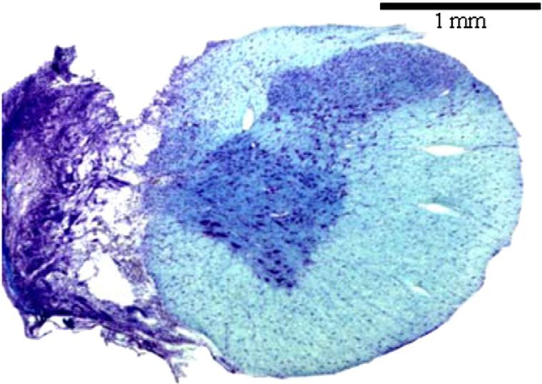
Fig. 1.
A representative histological section depicting a C2HS injury. This section was taken from cervical segment C2 at 8-week post-injury. The tissue was stained with luxol fast blue and cresyl violet. The absence of healthy tissue in the ipsilateral spinal cord suggests an anatomically complete C2HS.

