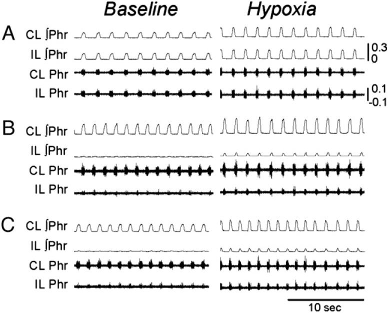
Fig. 6.
Examples of phrenic motor output recorded in anesthetized rats. Panels A-C present ipsilateral (IL) and contralateral (CL) phrenic neurograms recorded in control (panel A) and C2HS rats at 2- (panel B) and 8- (panel C) week post-injury during quiet breathing (baseline) and respiratory stimulation with hypoxia. Note that the IL and CL phrenic neurograms are similar in the control rat, but IL burst amplitude is dramatically reduced (vs. CL) at both 2- and 8-week post-injury. All scaling are the same between panels A and C; the calibration bars depict volts. ∫ indicates moving averaged or “integrated” neurogram.

