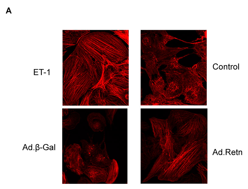
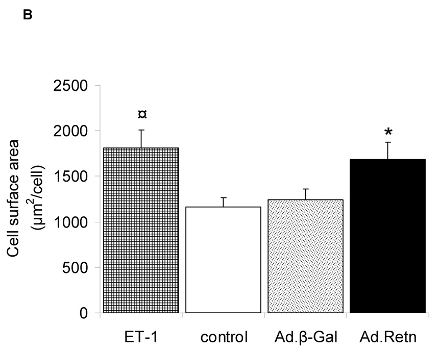
Figure 3. Effect of resistin on sarcomere organization and cell surface area.
A, Uninfected neonatal cardiomyocytes (control) or myocytes infected with Ad.β-Gal or Ad.Retn were incubated for 48 hours before staining with Alexa 594-conjugated phallodin. Endothelin-1 (ET-1), a hypertrophic agonist used as a positive control, induced highly organized sarcomere structures. Likewise, Ad.Retn-infected myocytes exhibit a large number of cells with highly organized sarcomeres compared with control or Ad.β-Gal-infected cells. At least 200 cells per condition were scored for the presence of highly organized sarcomeres. B, Cell surface areas of more than 100 individualized cells per condition from 3 independent experiments were measured using Image J software. ¤P<0.01 ET vs control; *P<0.01 Ad.Retn vs Ad.β-Gal. The mean values± SD are shown.


