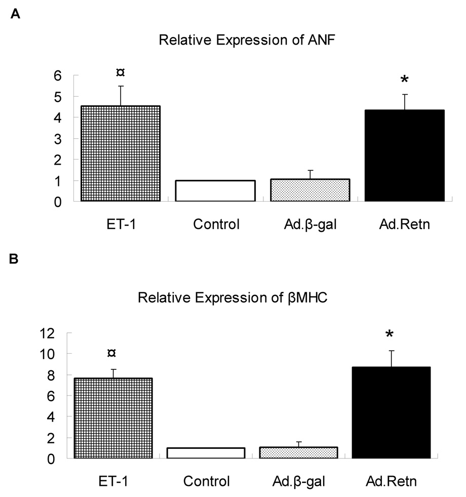
Figure 5. Induction of ANF and β-MHC by resistin.
Uninfected neonatal cardiomyocytes (control) or myocytes infected with Ad.β-Gal or Ad.Retn were incubated for 48 hours in serum-free media. The real time PCR for ANF and β-MHC was performed using primers specific to rat genes. The expression was normalized to 18S rRNA. Data normalized against control and expressed as fold change. ¤P<0.01 ET vs control; *P<0.01 Ad.Retn vs Ad.β-Gal‥

