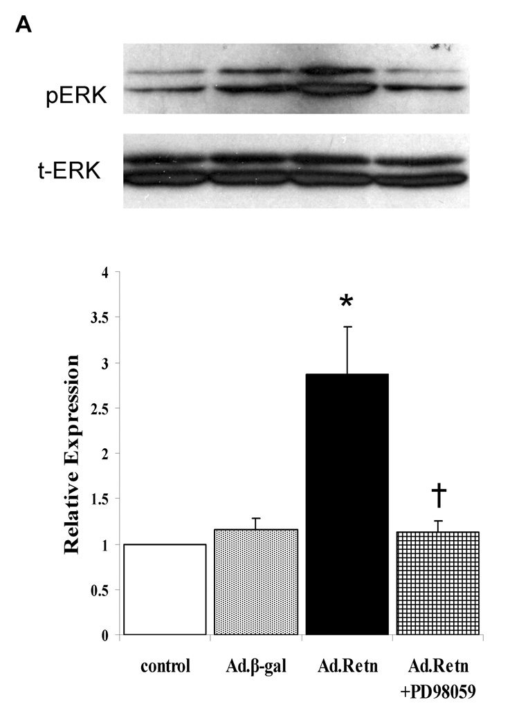
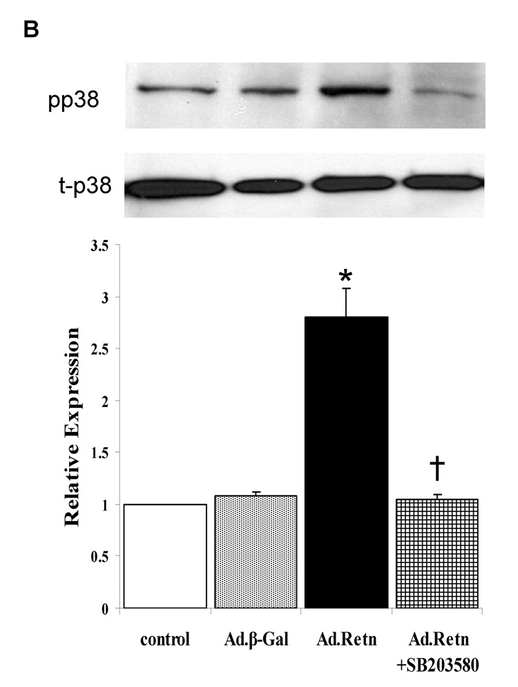
Figure 6. ERK and p38 MAP kinases are activated by resistin.
A and B, Uninfected neonatal cardiomyocyte (control) or myocytes infected with Ad.β-Gal or Ad.Retn were incubated for 24 hours in serum-free media. Cell lysates were matched for protein concentration and blotted onto PVDF membranes. The Blots were probed with phospho-specific antibodies against ERK and p38 and then reprobed for the corresponding total protein that confirmed equivalent loading of proteins. C and D, Quantitation of MAPKs activities. The intensity of each chemiluminescent band was quantified by densitometric scanning, and the activity of each MAPK was normalized against its corresponding total protein. Data are from at least three experiments. *P<0.05 Ad.Retn vs Ad.β-Gal; †P<0.05 Ad.Retn vs Ad.Retn + MAPK inhibitors.


