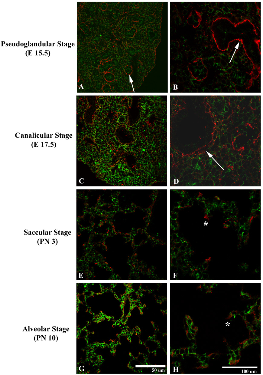
Figure 5.
Expression of uPARAP (green) and collagen I (red) during the pseudoglandular (A–B), cannalicular (C–D), saccular (E–F), and alveolar (G–H) stages. uPARAP demonstrates ubiquitous expression throughout the mesenchyme of the developing lung. Collagen I demonstrates few areas of mesenchymal staining. Collagen I is detected around developing airways (arrows) and, during later stages of development, along septal tips (*).

