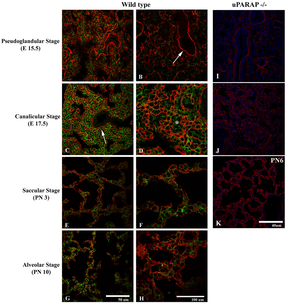
Figure 6.
Expression of uPARAP (green) and collagen IV (red) during the pseudoglandular (A–B), cannalicular (C–D), saccular (E–F), and alveolar (G–H) stages. Overlapping mesenchymal immunoreactivity for collagen IV and uPARAP is observed throughout all stages of development, particularly in the region of developing basement membranes (arrow). uPARAP expression, however, is detected in the absence of collagen IV immunoreactivity (*). I–K. Expression of collagen IV (red) in uPARAP −/− mice during pseudoglandular (I), cannalicular (J), alveolar (K) stages.

