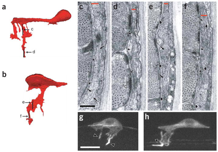
Figure 2.
HSN neurites are microtubule-containing, actin-rich structures. (a,b) HSN reconstructions based on serial EM sections, (a) Mid-L3 HSN with multiple neurites. (b) L3-L4 transition HSN with a single axon. Anterior is to the left and ventral is down, (c–f) Electron micrographs of HSN neurites. The position and orientation of the micrographs are indicated by the labeled vertical lines in a and b. In each section, a red bar marks the position of the HSN neurite. From left to right, each section includes muscle, basement membrane, the HSN neurite, epidermis and cuticle. Medial is to the left and ventral is down. In c and d, cross-sections of a neurite from a. (c) Proximal region showing microtubules in tangential view (arrows). Arrowheads span a stretch of basement membrane, (d) Distal region enriched in filaments (arrowheads). In e and f, cross-sections of a neurite from b. (e) Proximal region containing microtubules in tangential view (arrows) and ribosomes (fat arrows), (f) Distal region enriched in filaments (arrowheads) and ribosomes (fat arrows). In g and h, HSN neurons expressing GFP-actin at mid-L3 (g) and L3–L4 transition (h). Fluorescence is enriched in the distal regions of the neurites (arrowheads). Anterior is to the left and ventral is down. Scale bars: 200 nm in c–f; 5 μm in g,h.

