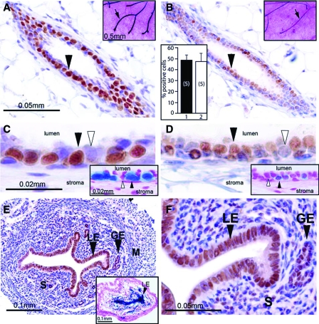
Figure 1.
Retention of mammary PR expression in the ovariectomized mouse. A, Immunohistochemical detection of PR is clearly evident in a transverse section of a mammary duct from an intact 8-wk-old wild-type virgin mouse (arrowhead). Inset shows robust β-gal activity (arrow) in a lacZ stained mammary whole mount from a similar aged intact PRlacZ knock-in mouse. B, Two weeks after ovariectomy, an 8-wk-old wild-type mouse exhibits a lower but detectable level of PR immunoreactivity in the mammary gland (arrowhead). Upper-right corner inset shows detectable β-gal activity (arrow) in a lacZ stained mammary gland whole mount from an ovariectomized 8-wk-old PRlacZ knock-in mouse. The histogram in the lower-left corner shows the percentage of mammary luminal epithelial cells positive for PR immunoreactivity in the intact and ovariectomized mouse (bars 1 and 2, respectively). The values represent the mean ± sem from five animals per group. C and D, Higher magnifications of regions shown in A and B, respectively. C, Note that robust PR immunoreactivity is restricted to the luminal epithelial compartment of the mammary gland (black arrowhead); cells scoring negative for PR expression are indicated by the white arrowhead. D, PR immunoreactivity is detected in the luminal epithelial compartment (black arrowhead); a subset of cells is negative for PR expression (white arrowhead). Insets in C and D show lacZ stained sections of mammary glands from the intact and ovariectomized PRlacZ knock-in mouse, respectively. Note the clear β-gal activity in both panels (black arrowhead); a cell negative for β-gal activity is indicated by a white arrowhead. E, Robust PR immunoreactivity is clearly detectable in the luminal and glandular epithelial (LE and GE, respectively) compartments of the uterus from an 8-wk-old ovariectomized mouse. Inset shows strong β-gal activity (black arrowhead) in the luminal epithelial compartment of a lacZ stained uterine transverse section from an 8-wk-old ovariectomized PRlacZ knock-in mouse. F, A higher magnification of a region shown in E; note the strong PR immunoreactivity in most cells of the luminal and glandular epithelial compartments (black arrowheads) and in a subset of stromal (S) cells. The luminal epithelial, glandular epithelial, stromal, and myometrial compartments are denoted by LE, GE, S, and M, respectively. Scale bars in A and C, apply to B and D, respectively; scale bars in insets in A and C apply to insets shown in B and D, respectively.

