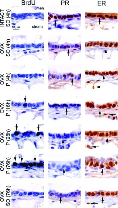
Figure 3.
Short progesterone exposure induces mammary epithelial proliferation in the ovariectomized mouse. High-magnification images of mammary gland sections from different hormone treatment groups [i.e. 8-wk-old intact or ovariectomized (OVX) wild-type mice treated with sesame oil (SO) or progesterone (P)] stained for BrdU incorporation, PR, and ER-α expression are shown as three sets of seven panels. Although the mammary epithelium of sesame oil-treated intact and ovariectomized mice does not exhibit BrdU incorporation, a significant increase in the number of luminal epithelial cells scoring positive for BrdU incorporation (arrow) in the mammary gland of the ovariectomized mouse is clearly evident by 16 h progesterone exposure, and this number significantly increases by 76 h progesterone exposure (arrows). Although ovariectomy significantly decreases the levels of mammary PR expression in the ovariectomized mouse [compare ovariectomized (sesame oil 4 h) with INTACT (sesame oil 4 h), and also see Fig. 1], mammary PR expression is further attenuated in the ovariectomized mouse when treated with progesterone for 16 h (arrow) and is absent by 76 h progesterone exposure (arrow). Unlike progesterone treatment, PR expression levels are not changed in the ovariectomized mouse with continuous sesame oil treatment for 76 h [arrow in ovariectomized (sesame oil 76 h)]. In contrast to PR expression, ER-α expression levels are not changed with progesterone exposure. ER-α expression levels in the luminal (vertical arrow) and stromal (horizontal arrow) compartments do not change in any hormone treatment group. Scale bar applies to all panels.

