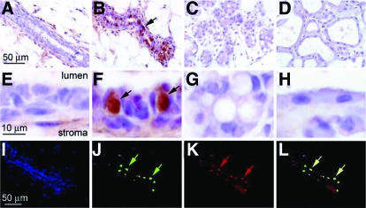
Figure 7.
Id4 expression is induced in the epithelial compartment of the murine mammary gland during early pregnancy and colocalizes with PR expression. A–D, Mammary gland sections immunohistochemically stained for Id4 expression from adult virgin (12 wk old), early pregnant (5.5 d post coitum), late pregnant (18 dpc), and lactating (d 7) mice, respectively. Although Id4 is not detected in the epithelial compartment of the mammary gland of the virgin, late pregnant or lactating mouse (A, C, and D, respectively), note the striking increase in Id4 expression in a subset of cells in the luminal epithelial compartment of the mammary gland of the early pregnant mouse [B (arrow)]. E–H represent higher magnification images of regions shown in A–D, respectively. Note the strong nuclear expression for Id4 in a subset of luminal epithelial cells of mammary gland at early pregnancy [F (arrows)]; approximately 30% of luminal epithelial cells score positive for Id4 expression. Dual immunofluorescence clearly demonstrates that Id4 and PR expression colocalize in the mammary epithelium of the early pregnant mouse. I, A 4′, 6′-diamidino-2-phenylindole-stained section of the mammary epithelium from an early pregnant (5.5 dpc) mouse. J, The same section as in I immunofluorescently stained for Id4. Note the punctate pattern for Id4 expression (green arrows), which agrees with the immunohistochemical data shown in B and F. K shows the same section immunofluorescently stained for PR expression; again note a similar punctate pattern for PR expression in this section (red arrows). Superimposing J and K confirms that Id4 and PR expression localize to identical cells (yellow arrows in L). Scale bar in A, E, and I apply to B–D, F–H, and J–L, respectively.

