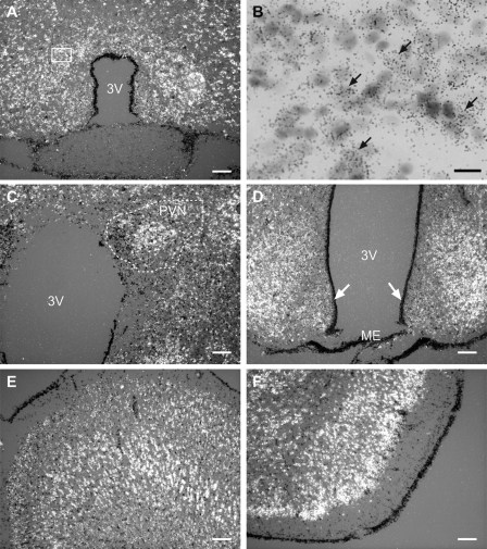
Figure 1.
KAP3 mRNA is expressed throughout the rat brain, as assessed by in situ hybridization using a [35S]UTP-labeled rat KAP3 cRNA probe and brain sections from 28- to 30-d-old late juvenile female rats. Selected regions are shown. A and B, AVPV of the POA; B, high-magnification view of A, depicting the presence of silver grain surrounding large, pale nuclei, characteristic of neurons, with examples denoted by arrows; C, periventricular nucleus (PVN) of the hypothalamus; D, VMH and ARC of the MBH; E, CTX; F, piriform cortex. Arrows point to tanycytes of the third ventricle (3V). Scale bars, 100 μm; bar in B, 20 μm.

