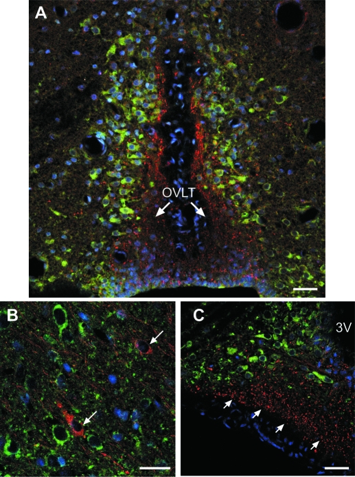
Figure 3.
KAP3 immunoreactivity is detected in cells near GnRH neurons but not in GnRH neurons themselves. The immunohistochemical reaction was performed on brain sections derived from two 28-d-old female rats. A, KAP3 immunoreactive neurons (green) adjacent to GnRH nerve terminals (red) projecting to the organum vasculosum of the lamina terminalis (OVLT) in the preoptic region of a 28-d-old female rat. B, KAP3 immunoreactive neurons (green) near GnRH neurons (red, denoted by arrows) lacking KAP3. C, KAP3 immunoreactivity is absent in GnRH nerve terminals of the ME (red) but is abundant in GnRH-negative neurons of the ARC (green). Cell nuclei stained with Hoescht are seen in blue. Scale bars, 10 μm.

