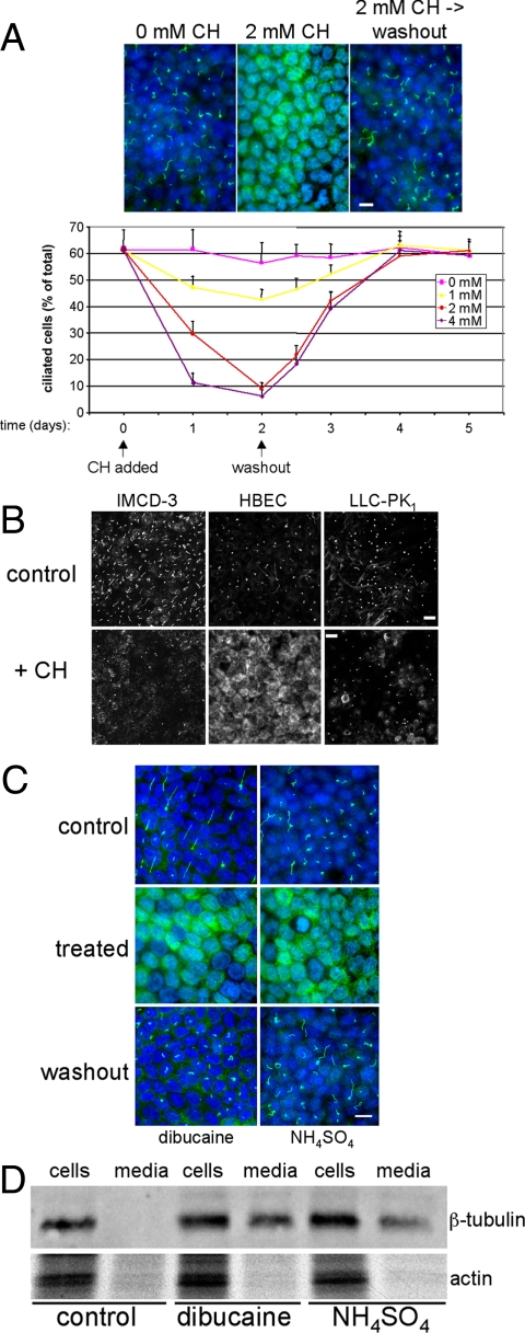
Figure 1.
Deciliation of polarized epithelial cells. (A) MDCK II cells were cultured in the absence or presence of indicated concentrations of CH for the durations specified. To examine the reversibility of observed effects, CH was removed at the indicated time (“washout”), and cells were cultured for an additional 3 d. At various time points, cultures were fixed and labeled with anti-acetylated tubulin antibodies (to mark primary cilia) and DAPI (to label nuclei). Bar, 10 μM. The percentage of ciliated cells was quantified by dividing the total number of cilia by the number of nuclei counted in 10 randomly selected fields from duplicate filters. Error bars, SEM (B) Polarized cultures of the indicated cell lines were incubated in the absence (control) or presence (+ CH) of either 2 mM (IMCD-3 cells and HBECs) or 6 mM (LLC-PK1 cells) CH for 3 d and then fixed and labeled with anti-acetylated tubulin antibodies. Bars, 20 μM. (C) Polarized MDCK II cells were cultured in the absence (control) or presence (treated) of either 0.75 mM dibucaine for 30 min or 30 mM NH4SO4 for 3 h. For washout, cultures were incubated an additional 48 h in the absence of deciliating agents. (D) Polarized MDCK II cells were cultured as in C, but in serum-free culture medium. Cell extracts and culture media samples, normalized for total protein content, were resolved by SDS-PAGE, and Western blots were probed for β-tubulin and actin. Note that tubulin, but not actin, is present in the culture medium after treatment of cells with dibucaine or NH4SO4, indicating the presence of shed cilia in the media.

