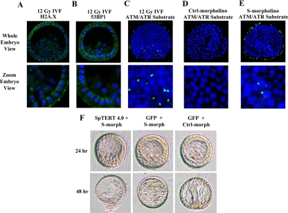
Figure 9.
Immunohistochemical analysis telomerase suppressed embryos and wt-SpTERT4.0-Lrescue phenotype. (A and B) Blastula-stage embryos were subjected to 12 Gy of radiation and subjected to gamma H2A.X or 53BP1 staining. No cross-reactivity with S. purpuratus was observed, and only nonspecific background was obtained. (C) Blastula-stage embryos were stained with an ATM/ATR phospho-substrate antibody (green) and DAPI counterstain (blue). Embryos were compressed at the time of mounting and visualization. Localization of the positive signal in the irradiated embryos was found to have a close, yet atypical association with the nucleus of the cell and possibly localized within the cytoplasm. (D) The nonirradiated control showed no positive staining. (E) The embryos injected with S-morpholino showed ATM/ATR substrate staining compared with controls. (F) Rescue attempt of telomerase suppressed embryos with wt-SpTERT4.0-L. Coinjection of wt-SpTERT4.0-L mRNA with the S-morpholino (that reduces telomerase activity) could not rescue the embryos. The control morpholino and GFP mRNA injected embryos showed no apparent aberration as it entered the gastrula stage of development.

