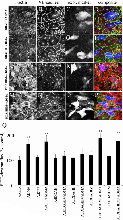
Figure 2.
DDAHI and DDAHII but not inactive DDAH mutants prevent ADMA-induced changes in the distribution of F-actin (A, E, I, and M), VE-cadherin (B, F, J, and N), and endothelial permeability (Q) in PAECs. (C, G, K, and O) Cells labeled with expression marker GFP; (D, H, L, and P) the corresponding composite images where F-actin is red, VE-cadherin is green, and GFP is blue (pseudocolors). Bar, 10 μm. DDAH and their inactive mutants were expressed via adenoviral gene transfer, and the cells were left untreated or were treated with 100 μM ADMA. * p < 0.05; ** p < 0.01, comparison with untreated controls, n = 5.

