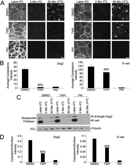
Figure 3.
MMP and EGFR inhibition interfere with the accumulation of an internalized pool of Dsg2. (A) SCC68 cells were cultured overnight with DMSO, TAPI, or PKI in 0.25 mM CaCl2. Dsg2 and E-cadherin (E-cad) monoclonal antibodies were used to measure internalization after 60 min. Bar, 20 μm. (B) Internalization was quantified by measuring the cytoplasmic fluorescence intensity using Metamorph Imaging. (C) SCC68 cells were incubated with DMSO, TAPI, or PKI overnight in the presence of 0.09 mM calcium, and cell surface biotinylation was performed as described to measure internalization after 60 min. (D) The ratio of cytoplasmic/surface intensity was used to measure internalization. PKI effectively blocked the accumulation of Dsg2 and E-cad, whereas TAPI was more effective inhibiting the appearance of internalized Dsg2.

