Figure 8.
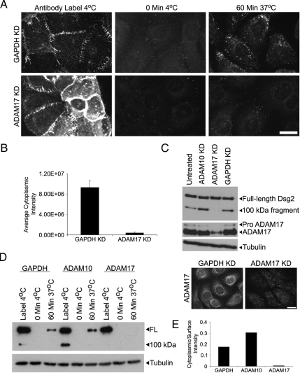
ADAM17 knockdown blocks the accumulation of an internalized pool of Dsg2. (A) SCC68 cells were transfected with GAPDH or ADAM17 siRNA in media containing 0.25 mM CaCl2 and analyzed 48 h after transfection. Cell surface Dsg2 was labeled with antibody, and internalization was assessed after 60 min at 37°C. ADAM17 localization was assessed using indirect immunofluorescence. Bar, 20 μm. (B) The average cytoplasmic intensity was measured using Metamorph Imaging. (C) Western blot analysis of Dsg2, ADAM17, and tubulin. ADAM17 was reduced by 65% percent under these conditions (C, top panel), and the residual ADAM17 exhibits a particulate pattern that appears distinct from Dsg2 staining (C, bottom right panel). (D and E) Cell surface biotinylation was performed to measure internalization after 60 min in GAPDH, ADAM10, or ADAM17 knockdown cells 96 h after transfection. Tubulin blot from RIPA lysates is shown to demonstrate equal starting material for all the samples. Cytoplasmic Dsg2 staining was absent in ADAM17 knockdown cells.

