Figure 7.
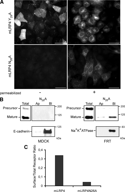
Mutations within the proximal NPxY motif of the LRP1 tail result in different receptor distributions. (A) FRT cells stably transfected with either mLRP4Y29A or mLRP4N26A were grown to confluence on coverslips and treated for indirect immunofluorescence with anti-HA under nonpermeabilized and permeabilized conditions, as indicated. Y29A distributed both at the apical cell surface (right panel) and intracellular vesicles (left panel), whereas N26A show only an intracellular localization, not being detectable at the cell surface. MDCK cells gave similar results (not shown). Scale bars, 10 μm. (B) Domain-specific cell surface biotinylation of MDCK and FRT cells expressing mLRP4N26A. In MDCK cells the protein was not detected at the cell surface. Controls confirmed the receptor expression (immunoblot of total lysate) and proper basolateral expression of E-cadherin. In FRT cells, however, a low amount of minireceptor was detected at the basolateral cell surface. Na+K+ ATPase was used as an endogenous basolateral marker. (C) MDCK cells expressing either mLRP4wt or the N26A mutant were consecutively incubated, either intact or after permeabilization with 0.1% saponin with an anti-HA and anti-mouse RPE-conjugated antibody, and analyzed by flow cytometry. The ratio of expression levels observed in intact versus permeabilized cells show 30% of the wild-type receptor versus no more than 4% of the N26A mutant at the cell surface.

