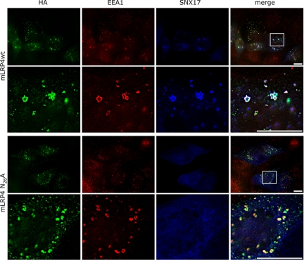
Figure 9.
Colocalization of minireceptors with endosomal markers EEA1 and SNX17. MDCK cells were microinjected with the plasmids encoding for RAP, SNX17-myc, and either the mLRP4wt or N26A minireceptors. Cells were processed for immunofluorescence to detect the HA-tagged minireceptor (green), the early endosome marker EEA1 (red), and the myc-tagged SNX17 (blue). In the amplified merged images, colocalization of the wild-type minireceptor with EEA1 in endosome-like structures was clearly visible; some of these structures also contain SNX17 (white dots). The N26A minireceptors practically did not colocalize with SNX17 and exhibited an increased colocalization with EEA1 (amplified merge image, yellow dots). Scale bars, 10 μm.

